Home Mammella Carcinoma mammario papillare

Carcinoma mammario papillare

Papillary Carcinoma of the Breast

Da dottvolpicelli

È un tipo di tumore molto raro (0,5-2% di tutte le neoplasie mammarie). È chiamato così perché al microscopio le cellule tumorali sono disposte in una forma che ricorda una felce. Si sviluppa di preferenza nelle donne di età >60 anni e in post-menopausa.

Il carcinoma mammario papillare può presentarsi in diversi tipi:

  • Carcinoma mammario papillare invasivo
  • Carcinoma mammario micropapillare invasivo
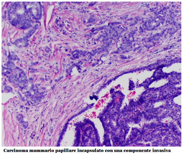
  • Carcinoma papillare intracistico/incapsulato/incistato
  • Carcinoma duttale papillare in situ

È spesso associato ad altri tipi di ca. mammario ed in particolare al carcinoma duttale in situ (DCIS).

EZIOLOGIA: Il carcinoma mammario papillare si verifica quando si sviluppano mutazioni nel DNA delle cellule dei dotti mammari.

DIAGNOSTICA:

  • All’esame obiettivo si palpa un nodulo o ispessimento del parenchima mammario e spesso sono presenti perdite ematiche dal capezzolo e introflessione del capezzolo.
  • Mammografia
  • Ultrasuonografia: è la modalità di imaging più ampiamente studiata. Si evidenziano tre profili ecografici di base: (1) massa intraduttale con o senza dilatazione duttale, (2) massa intracistica e (3) modello solido con una massa intraduttale che riempie completamente il dotto. Spesso sono presenti microcalcificazioni.
  • Biopsia centrale del nodulo usando un ago cavo per prelevare un campione di tessuto mammario da esaminare al microscopio 
  • Aspirazione con ago sottile (FNA) 

Immunoistochimica – il ca. papillare è  un cancro triplo negativo, (TNBC): sulle cellule tumorali non si riscontrano recettori per estrogeni (ER) nè recettori per il progesterone (PR) e non HER2. Quindi né la terapia ormonale né la terapia mirata sono efficaci. Il 10-20% di questi tumori possiedono recettori per gli androgeni (AR positivi) e da un punto di vista molecolare, questi tumori sono molto simili a quelli della prostata, e potrebbero quindi essere suscettibile agli stessi agenti terapeutici come la bicalutamide (farmaco antiandrogeno non steroideo).

Aspetto istologico: Il carcinoma papillare solido macroscopicamente appare come noduli ben circoscritti, densamente cellulari.  All’esame istologico si evidenziano proliferazioni cellulari che circondano i nuclei fibrovascolari, con o senza invasione.  La mucina intracellulare ed extracellulare associata è un reperto comune. I carcinomi papillari solidi sono spesso accompagnati da aree associate di carcinoma invasivo. 

TERAPIA

La chirurgia  è di solito il primo e il più importante trattamento per tutti i tipi di carcinoma papillare. Viene praticata una lumpectomia oppure mastectomia se il ca. è di notevole dimensioni oppure è interessata più di un’area mammaria. A volte è necessario un ulteriore intervento chirurgico se il margine del tessuto apparentemente normale che circonda il tumore che è stato rimosso durante la prima operazione non risulta assolutamente indenne all’esame istologico.

La chemioterapia post-chirurgica migliora la sopravvivenza a 5 anni (79% vs. 69%) ed attualmente è l’unica terapia medica effettivamente disponibile.


References:
  1.  Jemal A, Siegel R, Ward E, Hao Y, Xu J, Thun MJ. Cancer Statistics, 2009. CA A Cancer Journal for Clinicians. 2009;59(4):225–49. 
  2. Louwman MWJ, Vriezen M, Beek MWPMv, Nolthenius-Puylaert MCBJET, Sangen MJCvd, Roumen RM, et al. Uncommon breast tumors in perspective: Incidence, treatment and survival in the Netherlands. International Journal of Cancer. 2007;121(1):127–35. 
  3. McCulloch GL, Evans AJ, Yeoman L. Radiological features of papillary carcinoma of the breast. Clin Radiol. 1997;73:865–8.
  4. Grabowski J, Salzstein SL, Sadler GR, Blair S. Intracystic papillary carcinoma. Cancer. 2008;113(5):916–20. 
  5. Mulligan AM, O’Malley FP. Papillary lesions of the breast: a review. Adv Anat Pathol. 2007 Mar;14(2):108–19. 
  6. Collins LC, Schnitt SJ. Papillary lesions of the breast: selected diagnostic and management issues. Histopathology. 2008 Jan;52(1):20–9. 
  7. Ueng SH, Mezzetti T, Tavassoli FA. Papillary neoplasms of the breast: a review. Arch Pathol Lab Med. 2009 Jun;133(6):893–907. 
  8. Page DL, Salhany KE, Jensen RA, Dupont WD. Subsequent breast carcinoma risk after biopsy with atypia in a breast papilloma. Cancer. 1996 Jul 15;78(2):258–66.
  9. Hill CB, Yeh IT. Myoepithelial Cell Staining Patterns of Papillary Breast Lesions. American Journal of Clinical Pathology. 2005;123(1):36–44. 
  10. Collins LC, Carlo VP, Hwang H, Barry TS, Gown AM, Schnitt SJ. Intracystic papillary carcinomas of the breast: a reevaluation using a panel of myoepithelial cell markers. Am J Surg Pathol. 2006 Aug;30(8):1002–7. 
  11. Esposito NN, Dabbs DJ, Bhargava R. Are encapsulated papillary carcinomas of the breast in situ or invasive? A basement membrane study of 27 cases. Am J Clin Pathol. 2009 Feb;131(2):228–42.
  12. Maluf HM, Koerner FC. Solid papillary carcinoma of the breast. A form of intraductal carcinoma with endocrine differentiation frequently associated with mucinous carcinoma. Am J Surg Pathol. 1995 Nov;19(11):1237–44. 
  13. Nassar H, Qureshi H, Volkanadsay N, Visscher D. Clinicopathologic analysis of solid papillary carcinoma of the breast and associated invasive carcinomas. Am J Surg Pathol. 2006 Apr;30(4):501–7. 
  14. Moritani S, Ichihara S, Kushima R, Okabe H, Bamba M, Kobayashi T, et al. Myoepithelial cells in solid variant of intraductal papillary carcinoma of the breast: a potential diagnostic pitfall and a proposal of an immunohistochemical panel in the differential diagnosis with intraductal papilloma with usual ductal hyperplasia. Virchows Archiv. 2007;450(5):539–47. 
  15. Nicolas MM, Wu Y, Middleton LP, Gilcrease MZ. Loss of myoepithelium is variable in solid papillary carcinoma of the breast. Histopathology. 2007;51(5):657–65. 
  16. Fisher ER, Palekar AS, Redmond C. Pathologic findings from the National Surgical Adjuvant Breast Project (protocol no.4) VI. Invasive papillary cancer. Am J Clin Pathol. 1980;73:313–22.
  17. Walsh MM, Bleiweiss IJ. Invasive micropapillary carcinoma of the breast: Eighty cases of an underrecognized entity. Human pathology. 2001;32(6):583–9.
  18. Nassar H, Wallis T, Andea A, Dey J, Adsay V, Visscher D. Clinicopathologic Analysis of Invasive Micropapillary Differentiation in Breast Carcinoma. Mod Pathol. 14(9):836–41. 
  19. Pettinato G, Manivel CJ, Panico L, Sparano L, Petrella G. Invasive Micropapillary Carcinoma of the Breast. American Journal of Clinical Pathology. 2004 June;121(6):857–66. 
  20. Shah VI, Flowers CI, Douglas-Jones AG, Dallimore NS, Rashid M. Immunohistochemistry increases the accuracy of diagnosis of benign papillary lesions in breast core needle biopsy specimens. Histopathology. 2006 May;48(6):683–91. 
  21. Grin A, O’Malley FP, Mulligan AM. Cytokeratin 5 and estrogen receptor immunohistochemistry as a useful adjunct in identifying atypical papillary lesions on breast needle core biopsy. Am J Surg Pathol. 2009 Nov;33(11):1615–23. 
  22. Rabban JT, Koerner FC, Lerwill MF. Solid papillary ductal carcinoma in situ versus usual ductal hyperplasia in the breast: a potentially difficult distinction resolved by cytokeratin 5/6. Hum Pathol. 2006 Jul;37(7):787–93.
  23. Tan PH, Aw MY, Yip G, Bay BH, Sii LH, Murugaya S, et al. Cytokeratins in papillary lesions of the breast: is there a role in distinguishing intraductal papilloma from papillary ductal carcinoma in situ? Am J Surg Pathol. 2005 May;29(5):625–32. 
  24. Grin A, O’Malley FP, Mulligan AM. Cytokeratin 5 and Estrogen Receptor Immunohistochemistry as a Useful Adjunct in Identifying Atypical Papillary Lesions on Breast Needle Core Biopsy. The American Journal of Surgical Pathology. 2009;33(11):1615–23. doi: 10.097/PAS.0b013e3181aec446.
  25. Cristofano CD, Mrad K, Zavaglia K, Bertacca G, Aretini P, Cipollini G, et al. Papillary lesions of the breast: a molecular progression? Breast Cancer Research and Treatment. 2005;90(1):71–6. 
  26. Tsuda H, Takarabe T, Inazawa J, Hirohashi S. Detection of Numerical Alterations of Chromosomes 3, 7, 17 and X in Low-grade Intracystic Papillary Tumors of the Breast by Multi-color Fluorescence In Situ Hybridization. Breast Cancer. 1997 Dec;4(4):247–52. 
  27. Ganesan S, Karthik G, Joshi M, Damodaran V. Ultrasound spectrum in intraductal papillary neoplasms of breast. Br J Radiol. 2006 October 1;79(946):843–9. 
  28. Kim TH, Kang DK, Kim SY, Lee EJ, Jung YS, Yim H. Sonographic Differentiation of Benign and Malignant Papillary Lesions of the Breast. J Ultrasound Med. 2008 January 1;27(1):75–82.
  29. Lam WWM, Chu WCW, Tang APY, Tse G, Ma TKF. Role of Radiologic Features in the Management of Papillary Lesions of the Breast. Am J Roentgenol. 2006 May 1;186(5):1322–7. 
  30. Linda A, Londero V, Mazzarella F, Zuiani C, Bazzocchi M. Rare breast neoplasms: is there any peculiar feature on magnetic resonance mammography? La Radiologia Medica. 2007;112(6):850–62.
  31. Bernik SF, Troob S, Ying BL, Simpson SA, Axelrod DM, Siegel B, et al. Papillary lesions of the breast diagnosed by core needle biopsy: 71 cases with surgical follow-up. American journal of surgery. 2009;197(4):473–8. 
  32. Anderson W, SSD In situ male breast carcinoma in the Surveillance, Epidemiology, and End Results database of the National Cancer Institute. Cancer. 2005 Oct 15;104(8):1733–41.
  33. Burga A, Fadare O, Lininger R, Tavassoli F. Invasive carcinomas of the male breast: a morphologic study of the distribution of histologic subtypes and metastatic patterns in 778 cases. Virchows Archiv. 2006;449(5):507–12.
  34. Anan H, Okazaki M, Fujimitsu R, Hamada Y, Sakata N, Nanbu M. Intracystic papillary carcinoma in the male breast. A case report. Acta Radiol. 2000;41(3):227–9. 
  35. Bhatia A, Saikia Uma Nahar, Kumar Y. Rare Coexistence of Invasive Papillary Carcinoma with Infiltrating Ductal Carcinoma in Male Breast: Report of a Case. International Journal of Surgical Pathology. 2008 July 1;16(3):311–3.
  36. Blaumeiser B, Tjalma W, Verslegers I, et al. Invasive papillary carcinoma of the male breast. European Radiology. 2002;12(9):2207–10. 
  37. Dragoumis D, Tsiftsoglou A. Intracystic papillary carcinoma associated with ductal carcinoma in situ in a male breast. Journal of Postgraduate Medicine. 2008;54(1):39–40. 
  38. Imoto S, Hasebe T. Intracystic papillary carcinoma of the breast in male: case report and review of the Japanese literature. Jpn J Clin Oncol. 1998;28(8):517–20. 
  39. Pacelli A, Bock BJ, Jensen EA, Heerden JA, Reynolds C. Intracystic papillary carcinoma of the breast in a male patient diagnosed by ultrasound-guided core biopsy: a case report. Breast J. 2002 Nov–Dec;8(6):387–90. 
  40. Takayuki K, Takashi F, Eriko I, Miyuki T, Sadako A-T, Tadashi H. Intracystic papillary carcinoma of the breast in a male patient diagnosed by core needle biopsy: a case report. Breast (Edinburgh, Scotland) 2005;14(4):322–4.
  41. Tochika N, Takano A, Yoshimoto T, Tanaka J, Sugimoto T, Kobayashi M, et al. Intracystic carcinoma of the male breast: report of a case. Surg Today. 2001;31(9):806–9. 
  42. Zeppa P, Mascolo M, Zabatta A, Finelli L, Vetrani A, Palombini L. Aggressive papillary male breast carcinoma on fine-needle cytology sample. Diagn Cytopathol. 2003;29(6):360–3. 
  43. Solorzano CC, Middleton LP, Hunt KK, Mirza N, Meric F, Kuerer HM, et al. Treatment and outcome of patients with intracystic papillary carcinoma of the breast. American journal of surgery. 2002;184(4):364–8. 
  44. Fayanju OM, Ritter J, Gillanders WE, Eberlein TJ, Dietz JR, Aft R, et al. Therapeutic management of intracystic papillary carcinoma of the breast: the roles of radiation and endocrine therapy. American journal of surgery. 2007;194(4):497–500. 
  45. Baehner FL, Watson D, Ballard JT, Palmer G, Shak S. Gene expression by standardized quantitative RT-PCR in the special histologic subtypes of estrogen receptor positive invasive breast cancer. Breast Cancer Res Treat. 2008 [Abstr 3028] 
  46. Welsch C, Brown C, Goodrich-Smith M, Van J, Denenberg B, Anderson T, et al. Inhibition of mammary tumorigenesis in carcinogen-treated Lewis rats by suppression of prolactin secretion. J Natl Cancer Inst. 1979;63:1121 – 4. 


Potrebbe piacerti anche

Lascia il tuo commento

Inserisci la somma corretta Limite di tempo superato. Si prega di completare nuovamente il captcha.

Il Fertilitycenter.it è un sito informativo al fianco delle coppie per sostenerle ed informarle nel desiderio più grande, quello di avere un figlio.

ULTIMI ARTICOLI

fertilitycenter.it © 2023 All right reserved.

Questo sito Web utilizza i cookie per migliorare la tua esperienza. Supponiamo che tu sia d'accordo con questo, ma puoi annullare l'iscrizione se lo desideri. Accetto Leggi

-
00:00
00:00
Update Required Flash plugin
-
00:00
00:00